URETER ANATOMY

Gross Appearance
Length: 25 - 30 cm, in direct
relation to the height of the individual.
smooth S curve
Carries urine from renal pelvis to
bladder
Dividions

According to sacroiliac joint: Upper
1/3, Middle 1/3 (bet. Upper & lower sacroiliac j.) & Lower (1/3)
Areas of relative narrowing

(1) UPJ,
(2) crossing over iliac vessels,
(3) intramural part
Histology
Mucosa: transitional epithelium over lamina
propria (loose connective and elastic tissue).
Musculosa:
Upper part: run helically not arranged in
definite layers (considered inner long.& outer circ.)
lower part: longitudinal ms layer. & At UVJ,
trigonal ms reflect onto ureter
Waldeyer’s sheath: reflected trigonal ms onto lower 2-3
com of ureter
thin loose connective tissue at UVJ
separate Wldeyer’s sheath from long. ureteric ms
adventitia fibrous connective tissue. Peri-ureteric sheath is
thickened coat around ureter from intermediate stratum of retroperitoneum.
Relations : As followed from above downward,
Peritoneal relations
Abdominal ureter: lie within
retroperitoneum (intermediate stratum)
Pelvic ureter: covered by the
posterior peritoneum; lowermost part is closely attached to it, while the
juxtavesical part is embedded in vascular retroperitoneal fat.
Course
lies on the psoas muscles, on
transverse process L3,4,5
crosses common iliac artery
bifurcation (at pelvic brim)
passes medially to the sacroiliac
joints,
swings laterally near the ischial
spines
crossed by sup. vesical artery
(continue as obliterated umbilical a.) before entering bladder
pass medially to base of the bladder.
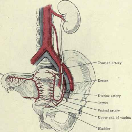
Structures that cross over ureter
In both sexes, sup. vesical artery crosses over
ureter before it enters bladder
In females, uterine arteries cross over juxtavesical
part of the ureters.
In males,
vas crosses anterior to the ureters over the lateral pelvic walls (after
it leave the internal inguinal rings) → lie medial to ureter before joining
S.V.

Blood Supply (mostly longitudinal)
[ARTEC]
·
abdominal ureter receives arterial branches from a medial
direction → dissect laterally
·
pelvic ureter receives arterial branches from a lateral
direction → dissect medially

A. Arterial
upper ureters → from renal arteries
mid ureter → from gonadal arteries.
Lower ureter → aorta (near
bifurcation), common
iliac, internal iliac (hypogastric), and sup. & inf. vesical arteries +
uterine a. (in females).
B. Venous
The veins are paired with the
arteries.
Nerve Supply

(autonomic) (unclear role)
From Inf. Mesenteric, testicular &
pelvic plexus which receive:
Ø
Sympathetic
from T11 – L2
Ø
Parasympathetic
from S2 –S4
Pacemaker of ureteric prestalsis lies
in renal pelvis, however, main stimulus for peristalsis is urine bolus
Lymphatic drainage
accompany bl.supply
upper ureter: Lumbar L.N. (Rt → interaortocaval, precaval & Lt
→ paraaortic),
middle ureter → common iliac L.N.s
lower urete → vesical, internal iliac & ext.
iliac L.N.s